Hip Arthritis
What is hip arthritis?
Hip arthritis involves the loss of cartilage at the end of the bones that make up the hip joint. This breakdown in the cartilage leads to pain, decreased range of motion of the hip, and difficulty performing daily activities.
What is the basic anatomy of the hip?
The hip is a ball-and-socket joint. The round ball is the femoral head which is at the top of the femur (thigh bone). The socket is the acetabulum which is part of the pelvis. The labrum is a special piece of cartilage that deepens the hip socket. The joint capsule and ligaments keep the femoral head in the correct position within the acetabulum.
What happens to the normal anatomy as hip arthritis develops?
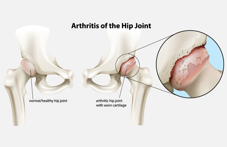
First, the cartilage thins out and starts to break down. If there is bone to bone contact, this can be very painful. The body tries to stabilize the joint and actually creates more bone called osteophytes, or bone spurs. These bone spurs reduce the normal range of motion of the hip. Lastly, the muscles surrounding the hip become very tight due to this lack of normal motion. Over time, abnormal hip mechanics can start to cause problems with back pain and knee pain.
What are the symptoms of hip arthritis?
- Pain: The classic location for hip arthritis pain is actually in the groin. Pain on the outside part of the hip is frequently associated with hip bursitis, or soft tissue inflammation. Pain in the buttock region is common with low back issues but can be related to the hip joint.
- Stiffness, Reduced Range of Motion: As hip arthritis progresses, the hip will lose range of motion. Patients will frequently report difficulty with putting shoes on.
- Difficulty with walking: Often times patients with hip arthritis have activity-related pain. Patients will report ability to walk only a short distance due to pain. Patients often have difficulty getting up from a seated position as well.
How do you make the diagnosis of hip arthritis?
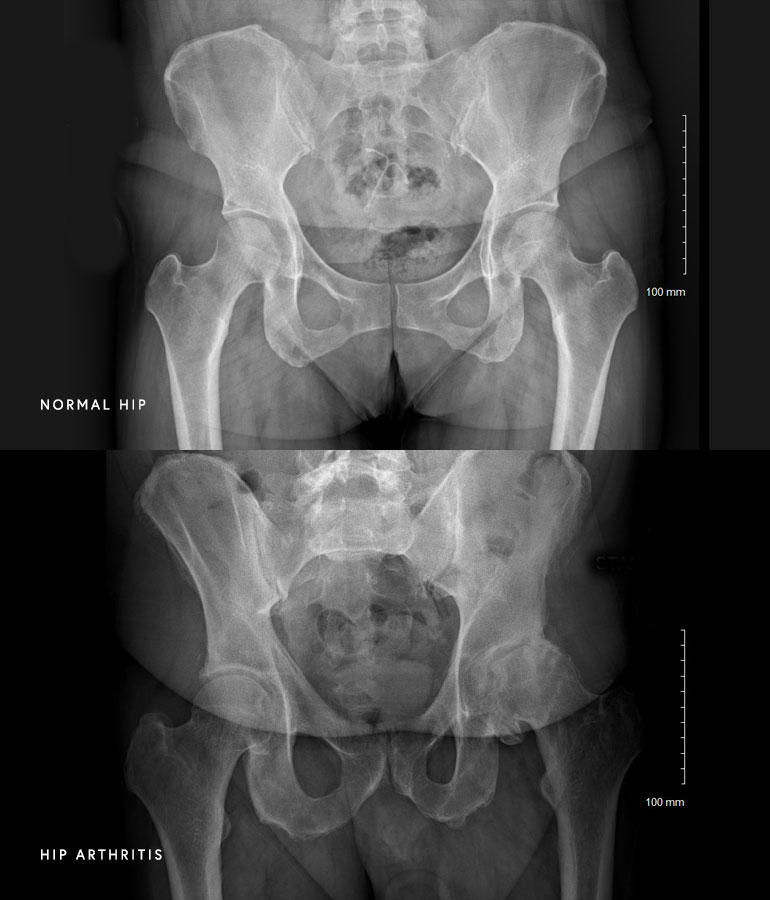
Dr. Kelly performs a thorough clinical evaluation on each patient that includes three major parts: history, physical examination, and imaging. The history focuses on certain activities that aggravate the patient’s symptoms as well as the location of pain. Dr. Kelly performs a physical examination that involves evaluating range of motion of the affected hip compared to the other hip. Additionally, he will ask the patient to perform various movements to try to determine if the pain is coming from the hip or the back. Finally, x-rays are reviewed which provide additional information about the shape and health of the hip joint. Sometimes additional studies are required such as MRI or x-rays of the lumbar spine.
What are common causes of hip pain?
It is important to make an accurate diagnosis of hip pain before offering treatment options. The most common causes for hip pain are reviewed below.
- Hip Arthritis: This is wear and tear disease of the cartilage in the hip joint. This typically presents with groin pain and decreased range of motion of the hip.
- Greater Trochanteric Bursitis: This is a very common cause of pain around the hip. This typically presents with pain on the lateral (outside) part of the hip. Patients frequently describe difficulty lying on their side. Treatment includes physical therapy and injections, but almost never surgery.
- Lumbar Spine: A pinched nerve in the lumbar spine can cause pain in the hip region without associated back pain. Most commonly this is located in the buttock region and may travel down the leg. Patients are treated with physical therapy and given a referral to spine specialist.
What are the treatment options for hip arthritis?
- Non-operative treatment: In almost all scenarios, Dr. Kelly provides non-operative treatment options for a patient with hip arthritis. This typically includes a combination of anti-inflammatory medication, physical therapy, and lifestyle modifications. See “Non-operative Treatment of Hip Pain” for more details.
- Total Hip Replacement: Hip replacement is an excellent treatment for patients who fail non-operative management. In a hip replacement, all of the cartilage surfaces of the hip joint are removed and resurfaced using a metal and plastic implant.
At a Glance
Dr. Mick Kelly
- Board-Certified Orthopedic Surgeon
- Fellowship Training in Hip and Knee Replacement
- Author of medical publications and numerous textbook chapters
- Learn more